69-Inch Diameter Cyclotron
In the late 1960s and early 1970s the cyclotron was reconfigured to expand its capabilities for new fields of study.
Overview
In the mid-1960s, Lewis decided to dismantle the cyclotron and replace it with a 69-inch diameter accelerator to improve the facility’s versatility, beam intensity, and energy levels. Construction began in 1970 and was completed in 1972. Afterwards, the cyclotron continued some of its previous areas of study, including the groundbreaking production of the iodine-123 isotope. NASA’s cancellation of its nuclear program in 1973 prompted an effort to increase use of the cyclotron for external customers.
Redesign of the Cyclotron
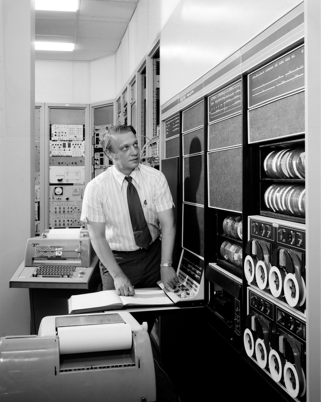
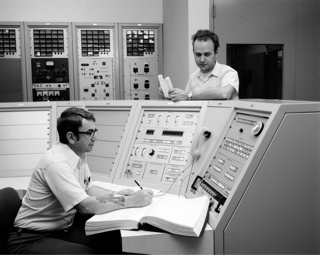
In 1965, members of the Lewis Radiation Physics Branch decided to reconfigure the center’s 60-inch cyclotron into an azimuthally—the angular distance between the particle’s direction and a fixed point—varying magnetic field (AVF) accelerator. This would provide the ability to accelerate a variety of particles over continuously variable energy ranges up to 50 million electron volts. It would also be one of the first cyclotrons with computerized controls for both the machine and experiment.
After the difficulties getting the existing accelerator up and running in the 1950s, researchers decided to essentially copy the design of a successful 50 MeV cyclotron at Michigan State University (MSU). Though larger, the new machine would have to fit into the Cyclotron Facility and utilize the existing steel yoke, magnetic coils, and power supplies.
In 1965, NASA hired William Brobeck to determine the cost and feasibility of the proposed modification, including three alternatives with enhanced capabilities. During his 20-year career at Ernest Lawrence’s University of California Radiation Lab, Brobeck had designed a series of cutting-edge, increasingly powerful cyclotrons. On September 3, 1965, Brobeck issued his report to Lewis and requested drawings of the MSU cyclotron to facilitate Lewis’ work.
The report offered plans to accomplish each of the alternatives, but stated the direct modelling of the MSU was the most cost efficient. The report also deemed any attempts to increase the power of the MSU design impractical and recommended against trying to incorporate new panels with sliding tuning.
Documents
- Cyclotron Redesign Request (1965)
- Cyclotron Redesign Proposal (1965)
- Cyclotron Conversion overview (1966)
- Cyclotron Upgrade Contracts (1968)
Installation
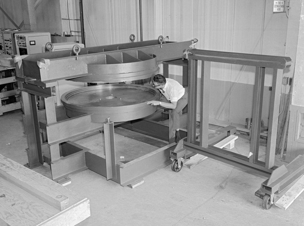
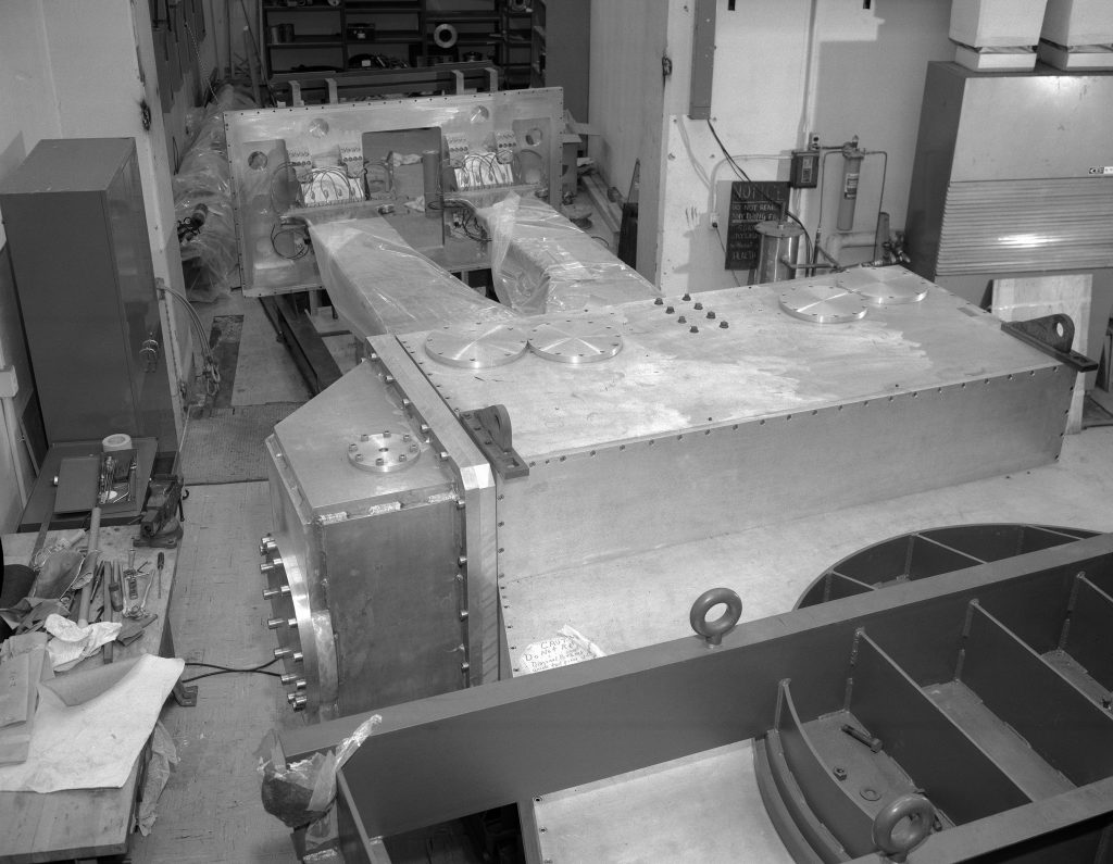
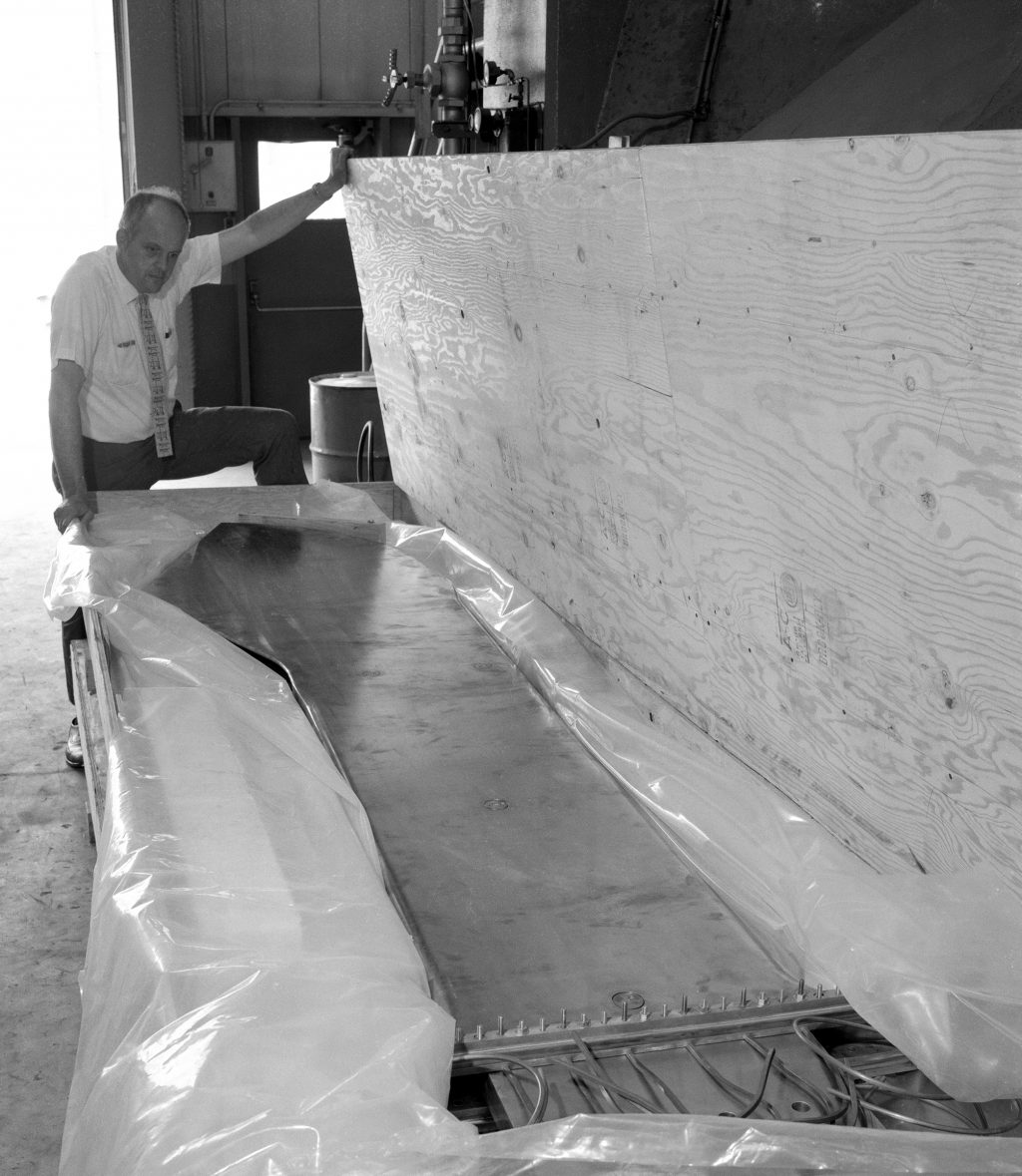
Congress approved funding for the upgrade in NASA’s fiscal year 1967 budget. The work was stretched out over 3 years for fiscal reasons. Activities began with a March 1968 review of the 60-inch cyclotron that found several deficiencies, including failures of the circuit monitoring the oscillator grid current. In June 1968, NASA contracted out the manufacturing of the new dees (hollow semicircular electrodes) and pole bases, and components began arriving the following summer.
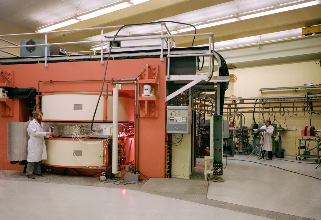
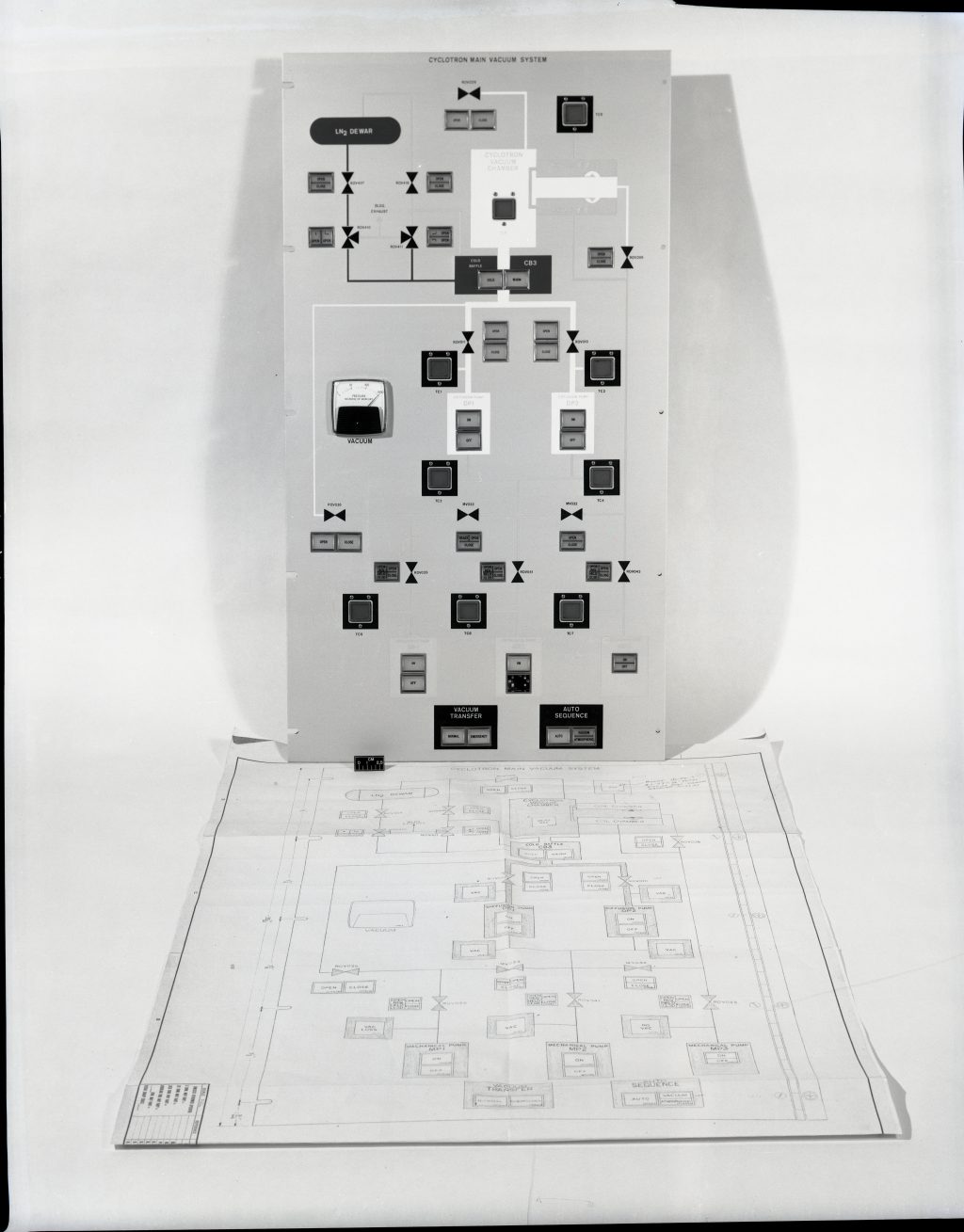
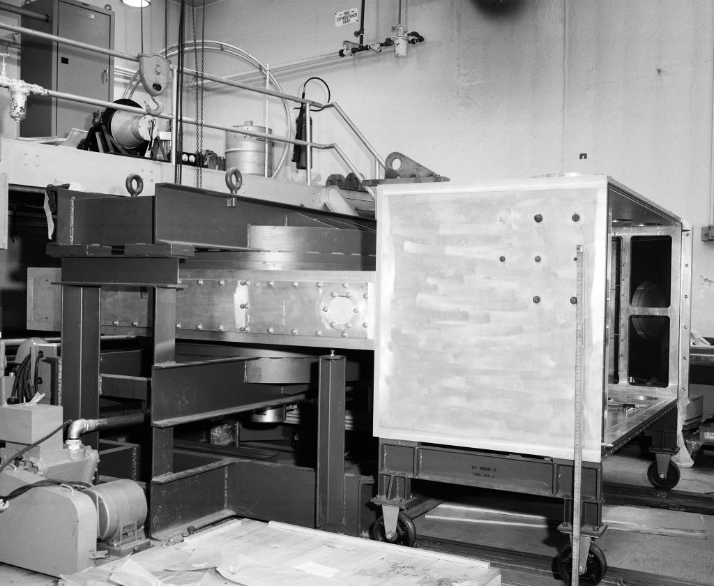
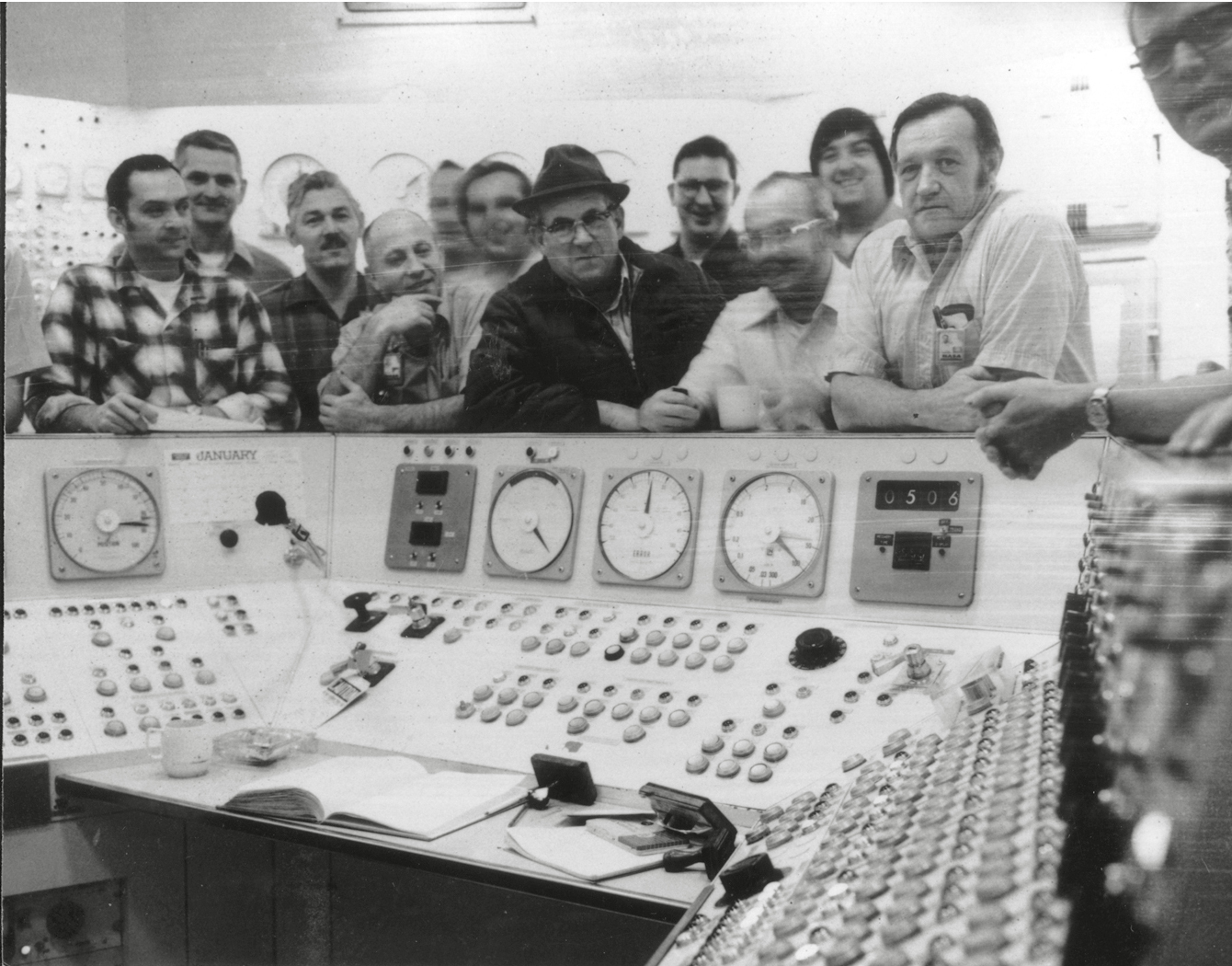
Researchers were able to continue using the existing 60-inch cyclotron until October 1970. In 1971, the old accelerator was disassembled, the new equipment installed, and initial magnetic mapping took place. Technicians completed the final wiring of the radio frequency (RF) power and vacuum systems. The control room in the Materials and Stresses Building basement was completely remodeled and upgraded. The new 69-inch cyclotron produced its first beam in 1972 and was conducting research in 1973.
The 69-inch cyclotron could accelerate proton (50 MeV), deuteron (28 MeV), helium-3 (87 MeV), and helium-4 (56 MeV) particles. Its varying energy field could be adjusted to all hydrogen and helium isotopes up to a maximum limit. Magnets directed the new device’s beam at a 90° angle so that an existing storage vault could be used as the beam room. The old beam room was used as a high resolution spectroscopy area.
External Customers
In January 1973, just as Lewis’ new 69-inch cyclotron was ready for testing, NASA terminated its nuclear propulsion and power programs. The final Apollo mission had just ended weeks before, and the agency was beginning the transition to the space shuttle. The cancellation resulted in the closure of Lewis’ Plum Brook Station [today, the Neil Armstrong Test Facility] and hundreds of layoffs. The center, however, decided to continue operation of the Cyclotron Facility.
The facility continued to support the activities of Lewis’ Materials and Stresses Division and Health Physics Office. It also contributed to other Lewis programs by continuing to test solar cell degradation, providing lead and gold anodes for use in NASA’s REDOX Project, and analyzing air samples for beryllium from the Global Air Sampling Program.
Without NASA’s funding for nuclear activities, however, the center had to reach out to external groups to fund projects for the cyclotron. During this period, the cyclotron also supported the following projects:
- Radiation effects on a Case Western Reserve University superconductor.
- Analysis of water samples for the Environmental Protection Agency and University of New Mexico.
- Calibration of space radiation detectors for Air Force Cambridge Research Laboratory.
- Supporting Langley Research Center’s materials damage research.
- Production of isotopes for Mount Sinai Hospital, the University of Indiana, and University of Cincinnati.
The University of Cincinnati used the isotopes for its experimental myocardial camera that was used to identify heart disease. In the 1970s researchers at the university developed the use of myocardial imaging to observe blood through a heart. The technique helps doctors identify heart disease and measure post-treatment improvements. The patient ingests a radioisotope that defines the damaged area for the camera. The Lewis cyclotron also created the thallium-201 radioisotopes used for the university’s early studies.
Nuclear Medicine Applications
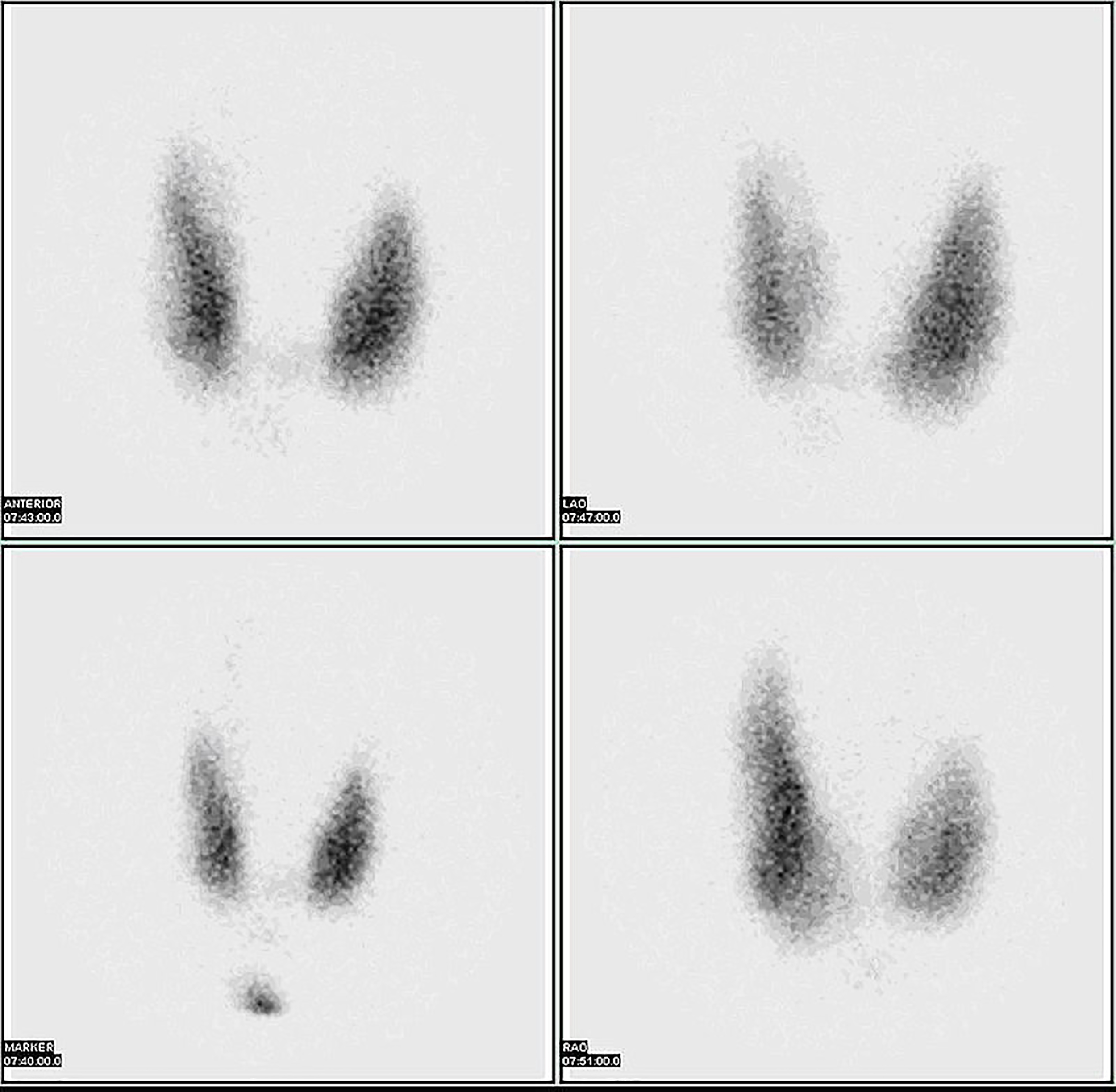
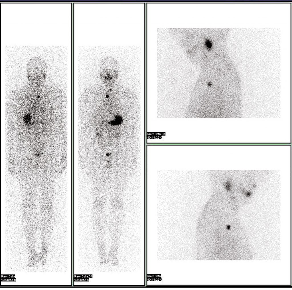
The term “nuclear medicine” refers to the use of radioactive substances to diagnose or treat health issues such as cancer, heart disease, and blood disorders. Radioisotopes are introduced into the patient and settle in organs or other desired locations. External gamma cameras record the radiation emitted by the isotope.
This imagery is useful in identifying abnormal growths and blockages of blood flow. Ernest Lawrence began experimenting with nuclear medicine in the 1930s. Its use to treat thyroid disease began in the late 1940s. By the 1970s, it was also being used to analyze other organs, and by the 1980s, it was used to detect heart disease.
In the late 1930s, U.S. researchers began experimenting using slightly radioactive iodine to diagnose and treat thyroid disease. The thyroid naturally accumulates iodine in its cells in order to generate hormones. Successful treatments using iodine-131, which destroys thyroid cells, occurred in 1941, and hundreds of treatments were applied in the post-War years. In 1951, the Food and Drug Administration approved iodine-131 patient use.
Iodine-131’s destruction of thyroid cells was useful for treating disease, but less than optimal for imaging applications. A purer form of iodine, iodine-123, was considered a better option for imaging because it provided clearer images and did not harm healthy thyroid cells. Iodine-123, however, was extremely difficult to produce.
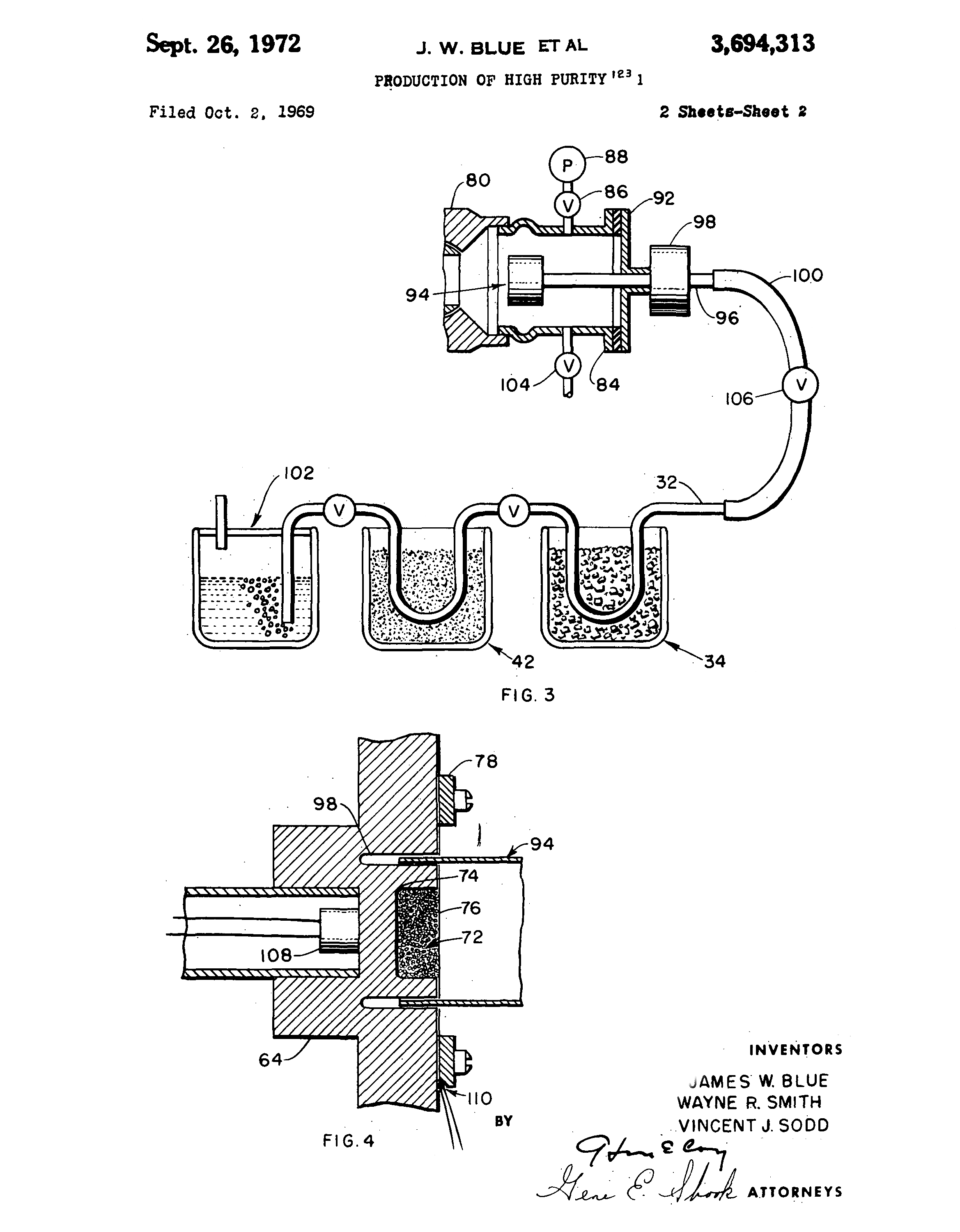
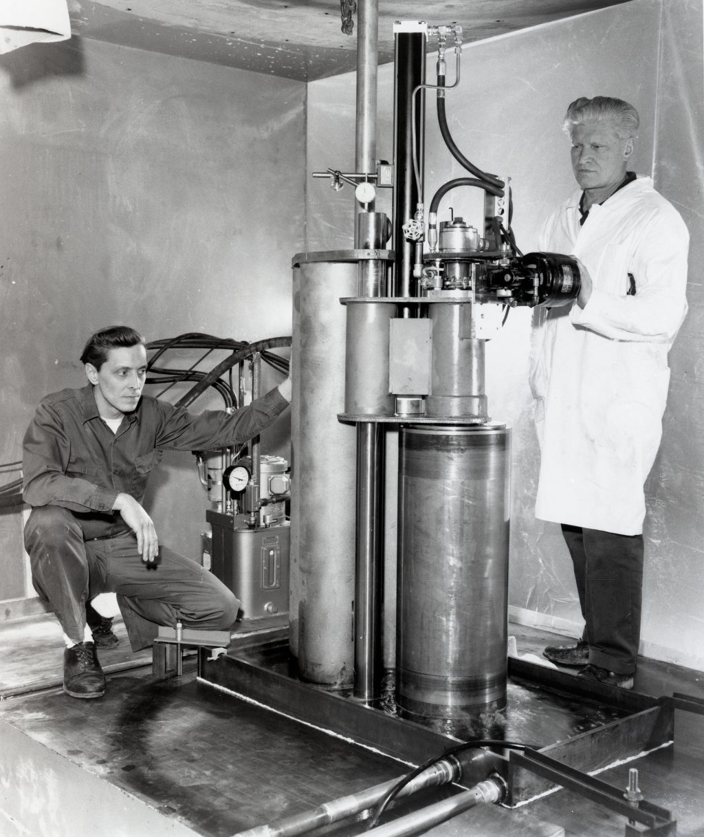
Researchers at other institutions attempted to create the isotope by producing xenon-123 and extracting the iodine-123 using chemicals or thermal cycling. These methods significantly reduced the yield, however. At Lewis, researcher James Blue worked with colleagues from the Nuclear Medicine Laboratory in Cincinnati to develop a production method in which tellurium-122 is bombarded to produce xenon-123, which decays into iodone-123.
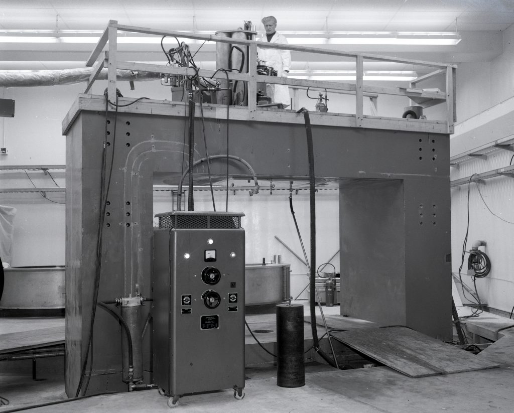
The Lewis cyclotron could generate several doses of iodine-123 in just hours. The key to the system was the use of cesium as both the working fluid and the target. Hence, there was a continuous cycling of the target material and extraction of the xenon-123, which decayed into iodine-123.
Today iodine-123 is the standard radioisotope for thyroid imaging. Compared to iodine-131, a much smaller quantity was required to produce clearer radiographs, and it stayed in the body for a significantly shorter period of time.
Documents
- Self-Vapor Cooled Targets for Production of I-123 (1974)
- Cyclotron Production of I-123 (1970)
- I 123 Production for Use in Nuclear Medicine (1971)